
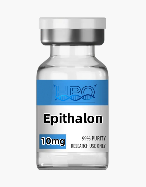
Epithalon Research Peptide: Identity, Evidence, and Laboratory Guide
FOR RESEARCH USE ONLY. Epithalon research peptide is supplied solely as an analytical and in vitro laboratory reagent. Not for human or veterinary use, food, cosmetic, household, diagnostic, or therapeutic purposes.
Overview
Epithalon research peptide is a sequence-defined, linear tetrapeptide composed of L-alanine, L-glutamic acid, L-aspartic acid, and glycine. Its sequence is L-Ala-L-Glu-L-Asp-Gly (AEDG), also written H-Ala-Glu-Asp-Gly-OH. For the standard neutral, unsolvated free peptide, authoritative identity records give the molecular formula C14H22N4O9, an average molecular mass of 390.35 g/mol, CAS Registry Number 307297-39-8, PubChem CID 219042, and FDA Unique Ingredient Identifier O65P17785G.[1][2] These identifiers describe a chemical structure; they do not establish the composition of an individual lot, regulatory approval, clinical utility, or suitability for a particular assay.
The spellings Epitalon and Epithalon usually refer to the same AEDG tetrapeptide in the scientific literature. Epithalone is another encountered spelling. By contrast, Epithalamin denotes an animal pineal-tissue peptide extract or peptide complex, not pure sequence-defined AEDG. Results obtained with that heterogeneous extract cannot be assigned automatically to synthetic Epithalon. Researchers should record the exact test article named in each paper and avoid merging these evidence streams.
Research interest in AEDG spans telomere biology, nucleic-acid and chromatin models, circadian readouts, redox-sensitive assays, cellular differentiation, immune signaling, and organismal survival studies. The evidence is predominantly preclinical, methodologically varied, and concentrated in a limited number of research groups. Several publications use transformed cell lines or historical animal models; some mechanistic conclusions rely on docking, fluorescence probes, or indirect biomarkers. This page therefore presents hypotheses and experimental controls rather than health claims. Chemical identity alone predicts none of the reported biological observations.
Molecular identity
| Catalog description | Epithalon research peptide |
|---|---|
| Common literature names | Epitalon; Epithalon; Epithalone; AEDG peptide; Ala-Glu-Asp-Gly |
| Defined sequence | L-Ala-L-Glu-L-Asp-Gly (AEDG) |
| Terminal form | H-Ala-Glu-Asp-Gly-OH; free amino and carboxyl termini |
| Architecture | Linear, non-cyclic, non-amidated tetrapeptide |
| Disulfide status | No disulfide bond; the sequence contains no cysteine |
| Free-peptide formula | C14H22N4O9 (neutral, unsolvated parent) |
| Average molecular mass | 390.35 g/mol for the standard free peptide |
| Monoisotopic mass | 390.138678 Da for the neutral free peptide |
| CAS Registry Number | 307297-39-8 for the standard free peptide; verify the declared form on the lot documents |
| PubChem | CID 219042 |
| FDA substance identifier | UNII O65P17785G; an identity code, not an approval |
| InChIKey | HGHOBRRUMWJWCU-FXQIFTODSA-N |
| Material represented by this entry | Lot-specific research reagent; the CoA and label control the actual salt, counterion, water, content, purity, and storage status |
| Intended use | Analytical, biochemical, and in vitro laboratory research only |
The formula, mass, CAS number, and UNII above apply to standard Epitalon free peptide. FDA materials distinguish that structure from Epitalon acetate, reported as H-Ala-Glu-Asp-Gly-OH·CH3CO2H, with CAS 307297-40-1 and a formula mass of 450.40 g/mol.[3] A lot described only by the common name cannot be assumed to be free peptide. Residual acetate, another counterion, water, or solvent can change gross mass and assay calculations even when the AEDG active moiety is present.
Names, source, and structural boundaries
Epitalon and Epithalon are transliteration variants rather than different peptide sequences when authors explicitly specify AEDG. The sequence declaration is more reliable than the common name. Epithalamin is materially different: it is described in older literature as a pineal peptide preparation derived from animal tissue. It may contain multiple peptides and other process-dependent constituents. A publication testing Epithalamin is not direct evidence for purified AEDG, and a synthetic AEDG lot should not be represented as an animal extract.
AEDG has three peptide bonds, three acidic side-chain or terminal carboxyl groups, a free N-terminus, and no aromatic amino-acid side chain. Its charge distribution varies with pH and ionic strength. The absence of cysteine means the native sequence cannot form an intramolecular or intermolecular disulfide bond. It also contains no methionine, tryptophan, or tyrosine. Those absences simplify some impurity questions but do not eliminate hydrolysis, stereochemical impurities, aggregation, adsorption, or sequence-related degradation.
The Asp-Gly junction deserves attention in stability-indicating methods because aspartyl sequences can undergo pathway-dependent rearrangement or cleavage. Intact mass alone may not separate an isomeric product from the intended sequence. The peptide also lacks a strong aromatic ultraviolet chromophore, so low-wavelength HPLC detection can be sensitive to solvent background, baseline drift, and integration settings. Its polarity can produce weak reversed-phase retention. Method development may therefore require qualified ion-pairing, hydrophilic-interaction, mixed-mode, or ion-exchange conditions, supported by orthogonal mass or structural data.
Seven research themes and their limits
1. Telomerase and telomere-maintenance models
A 2003 report found that AEDG exposure in telomerase-negative human fetal fibroblast cultures was associated with expression of the telomerase catalytic subunit, detectable enzyme activity, and telomere elongation.[4] The experiment is frequently cited, but a single cultured-cell system cannot establish a universal mechanism or organism-level outcome. Cell source, replicative history, culture stress, assay normalization, and telomere-method choice are important confounders.
A 2025 study examined normal epithelial and fibroblast cells alongside 21NT and BT474 breast cancer cell lines. It reported telomere-length changes associated with hTERT/telomerase readouts in normal cells and with alternative lengthening of telomeres (ALT)-related readouts in the transformed lines.[5] A correction was later published and should be read with the article.[6] The cancer-cell ALT observation is not a favorable product property; it is a model-specific result that complicates any simplistic narrative about telomere extension. It calls for direct assessment of transformation state, genomic instability, ALT markers, and proliferation. Telomere qPCR, hTERT transcript abundance, and enzyme assays answer different questions and should not be treated as interchangeable.
2. DNA, chromatin, and gene-expression hypotheses
Fluorescence studies using labeled short peptides reported intracellular and nuclear signal in HeLa cells and sequence-dependent quenching interactions between unmodified peptides and selected oligonucleotides.[7] Other publications described changes in heterochromatin-related cytological readouts in cultured lymphocytes.[8] These observations motivate binding and chromatin experiments, but they do not prove that native AEDG occupies a defined genomic site or directly controls transcription in intact cells. A fluorescent tag can change charge, transport, localization, and binding. Quenching can arise from several molecular processes, and computational complementarity is not a measured dissociation constant.
A rigorous program should distinguish cell entry, nucleic-acid association, histone association, chromatin accessibility, and functional transcription. Useful approaches include native-peptide competition, label-free biophysics, sequence-mutated oligonucleotides, chromatin-accessibility assays, promoter perturbation, and time-resolved transcript measurements. Docking can generate testable conformations but cannot substitute for occupancy, stoichiometry, kinetics, or causal perturbation.
3. Pineal, melatonin, and circadian readouts
An older study in female rhesus monkeys of different ages used enzyme immunoassays and reported changes in evening melatonin and cortisol rhythmicity after Epithalon exposure.[9] This is historical animal evidence, not a clinical conclusion. Circadian outputs are highly sensitive to lighting schedule, sampling density, season, feeding, handling stress, sex, and baseline phase. A single morning-versus-evening contrast cannot establish phase, amplitude, or period.
Laboratory follow-up should use prespecified zeitgeber conditions, frequent sampling across complete cycles, blinded sample analysis, and model-based rhythm statistics. Melatonin abundance, synthetic-enzyme expression, clock-gene phase, and behavioral activity are separate endpoints. The common name’s association with the pineal gland does not establish organ selectivity, and Epithalamin extract results must remain separate from sequence-defined AEDG results.
4. Oxidative-stress and mitochondrial readouts
Rat studies have reported changes in total antioxidant measures and enzyme activities in serum or tissues after exposure to pineal peptide preparations, including Epitalon.[10] A mouse-oocyte culture study reported changes in ROS-sensitive fluorescence, mitochondrial membrane-potential signal, mitochondrial DNA copy number, spindle features, and apoptosis-associated endpoints during post-ovulatory culture aging.[11] These are model-bound associations. They do not establish that AEDG is a direct radical scavenger or a general mitochondrial modulator.
Redox probes can be affected by probe loading, esterase activity, cell number, light, metal ions, and optical interference. Membrane-potential dyes are influenced by mitochondrial mass and dye handling. Stronger designs include cell-free interference controls, chemically distinct ROS methods, respiration or flux measurements, antioxidant-enzyme activity with protein normalization, and recovery of intact AEDG from the matrix. Report both null and adverse cellular readouts rather than collapsing them into a broad protective label.
5. Neural differentiation and retinal cell models
A 2020 study in human gingival mesenchymal stem cells reported changes in transcripts and proteins associated with neurogenic differentiation and paired these results with molecular modeling of AEDG interactions with histone H1 variants.[12] The experimental expression changes and the docking model are different evidence layers. Marker increases do not alone demonstrate mature neuronal identity, electrophysiological function, or direct histone binding.
A 2025 ARPE-19 high-glucose cell model reported changes in scratch-closure, ROS-sensitive signal, epithelial-to-mesenchymal-transition markers, and fibrosis-related transcripts.[13] Scratch closure may reflect migration, proliferation, survival, or image-segmentation choices. ARPE-19 cells under one culture condition do not reproduce the architecture, exposure barriers, or multicellular signaling of intact retina. Follow-up should separate migration from proliferation, include osmotic controls, verify cell identity, and confirm key findings in an orthogonal retinal model.
6. Immune signaling and proliferative endpoints
A mouse stress-model study evaluated thymocyte blast transformation and IL-1β-associated neutral sphingomyelinase activity, reporting condition-dependent changes with Epitalon.[14] A later THP-1 study compared several short peptides and peptide complexes in monocytic and macrophage-like states, observing peptide- and state-dependent signaling, proliferation, and cytokine readouts.[15] Neither design identifies a universal immune target for AEDG.
Immune assays should control differentiation state, passage, endotoxin, serum lot, cell density, and vehicle or counterion. Cytokine abundance, kinase phosphorylation, viability, and cell number should be measured separately. When several peptides are screened together, a class-wide result cannot be assigned automatically to Epithalon. Confirmatory work should predefine one primary endpoint and use pathway perturbation before proposing direct signaling.
7. Organismal survival and tumor-incidence experiments
A Drosophila study reported sex- and condition-dependent survival differences after AEDG was present during development, while also reporting a non-monotonic relationship across the tested concentrations.[16] A rat study under different illumination regimes reported no lifespan change under the standard light-dark condition and different results under natural or constant illumination; tumor-incidence findings also varied by lighting condition.[17] The null and context-dependent findings are as important as the positive ones.
These historical models do not establish an effect in people, do not validate a consumer application, and do not support cancer-related or lifespan marketing. Developmental exposure in flies and altered-light rodent paradigms have distinct biology and bias risks. Reproducibility work should use randomized allocation, blinded scoring, adequate cohort size, prespecified censoring, full survival curves, competing-risk analysis, pathology review, environmental monitoring, and replication by independent laboratories.
Appropriate laboratory applications
- LC–MS, HPLC, capillary-electrophoresis, or mixed-mode method development for a small acidic tetrapeptide;
- intact-mass, fragmentation, adduct, counterion, water, adsorption, and matrix-recovery studies;
- cell-free peptide–oligonucleotide or peptide–histone association experiments using orthogonal methods;
- in vitro telomerase, telomere-length, ALT-marker, chromatin-accessibility, and transcriptional assay development;
- in vitro circadian reporter, redox, mitochondrial, differentiation, migration, or immune-signaling studies with qualified controls; and
- stability-indicating analytical work in defined research buffers and culture matrices.
These examples are research categories, not validated protocols. Historical animal studies are summarized only to define the evidence base and its limitations; they do not change the intended use of this material.
Experimental design and interpretation
- Verify the test article. Record the lot number, declared chemical form, sequence, peptide content, counterion, water, purity method, storage history, and opening date. Confirm that the reference material and test article represent the same form.
- Predefine the biological question. Separate uptake, binding, hTERT expression, telomerase activity, telomere length, ALT activity, chromatin change, redox signal, and cell phenotype. Select a primary endpoint before data collection.
- Use informative in vitro controls. Depending on the question, include untreated, vehicle, counterion-matched, scrambled-sequence, reversed-sequence, and free-amino-acid-mixture controls. A method-positive control establishes assay competence but does not validate AEDG specificity.
- Build a concentration–time matrix. Select the experimental range from solubility, recovery, matrix integrity, cytotoxicity, and pilot signal. Do not transfer a nominal concentration between unrelated models without confirming actual exposure.
- Measure test-article integrity. Determine whether intact AEDG remains recoverable through the experimental interval. Proteases, pH, serum components, vessel binding, and temperature can change free concentration or generate fragments.
- Triangulate telomere biology. Pair a telomerase activity method with hTERT RNA or protein, and pair relative telomere qPCR with an orthogonal length method where feasible. If transformed cells are used, include C-circle, ALT-associated PML body, recombination, and growth-state readouts.
- Control assay interference. Run peptide-plus-reagent wells without cells for fluorescent, luminescent, colorimetric, and redox methods. Confirm central findings with a chemically different readout.
- Design for reproducibility. Use independent biological replicates, randomized plate positions, blinded imaging, documented exclusions, batch tracking, appropriate multiple-testing control, and complete reporting of cell source and passage.
- Test mechanism, not association alone. Establish temporal ordering, direct engagement, loss-of-function or blockade, and rescue logic. A docking score, transcript change, or correlated biomarker does not by itself show causality.
Analytical quality control
A credible release package separates identity, sequence, chromatographic purity, and net peptide content. An HPLC area result is method-specific and does not establish sequence, stereochemistry, total amount, water, counterion, or absence of non-UV-active impurities. No universal purity value should be inferred from the product name. Fit-for-purpose control of Epithalon research peptide may include:
- Identity: high-resolution LC–MS matched to the declared free peptide or salt form, with mass tolerance, isotope pattern, adduct assignments, and traceable raw data.
- Sequence and stereochemistry: justified MS/MS or comparison with a qualified reference. Chiral or enzymatic analysis may be necessary because intact mass cannot prove every L-configuration or distinguish all isomers.
- Related substances: a stability-indicating separation with stated column, mobile phases, gradient, detector settings, integration rules, reporting threshold, and system suitability. Demonstrate resolution of truncations, deletion sequences, Asp-Gly-related products, and process impurities where available.
- Net peptide content: quantitative amino-acid analysis, calibrated quantitative NMR, or a validated reference-standard assay. Report content separately from gross fill mass and chromatographic area purity.
- Water, counterion, and process residues: Karl Fischer analysis when applicable, ion chromatography for declared counterions, and solvent or elemental testing based on synthesis and purification knowledge.
- Aggregation and particulate assessment: use appropriate orthogonal methods when process knowledge or the intended assay makes these attributes relevant. A clear appearance does not exclude soluble aggregates or subvisible particles.
- Biological-assay compatibility: endotoxin, bioburden, or mycoplasma results only where required by the in vitro system. Such tests do not establish suitability outside research.
Because AEDG has no aromatic residue, low-wavelength UV response should be qualified for specificity and linearity. LC–MS methods should assess sodium, potassium, and solvent adducts rather than misassigning them as peptide variants. ICH Q2(R2) provides an official framework for analytical-procedure validation, including specificity, accuracy, precision, range, and robustness; voluntary use of that framework does not convert a research reagent into a regulated medicine.[18]
Lot-specific handling
The current lot label and CoA control handling. Verify the declared free-peptide or acetate form, peptide content, water, counterion, storage condition, transport history, and retest status before opening. Do not substitute generic web values for lot documentation. If the form is ambiguous, place the material on hold until identity and counterion status are resolved.
Handle dry material as a research chemical with incompletely characterized occupational hazards. Use trained personnel, task-appropriate protective equipment, controlled weighing practices, and the current safety data sheet. Minimize moisture exposure, uncontrolled light, and unnecessary temperature excursions. Allow the sealed container to reach the laboratory handling condition specified by the supplier before opening when condensation is a concern.
Any solution-phase analytical work should follow a laboratory-approved, matrix-specific SOP; this page intentionally provides no preparation procedure. Record the solvent or buffer, pH, container material, actual peptide content, and elapsed analytical interval. Low-binding vessels may be evaluated by recovery studies. Establish integrity in the exact matrix and container with a stability-indicating method, and avoid assigning a generic usable interval without lot- and method-specific data. Dispose of material and contaminated consumables through institutional chemical-waste procedures.
Evidence and regulatory boundary
The published record is exploratory and does not constitute a validated product specification. Many studies originate from overlapping investigators, sample sizes are sometimes limited, terminology is inconsistent, and old papers may not meet current reporting standards. Results span purified AEDG, material of incompletely stated form, and the distinct Epithalamin extract. Reproducibility across independent laboratories, chemically characterized lots, and orthogonal methods remains an important need.
PubChem CID 219042 and FDA UNII O65P17785G are substance identity records. A UNII is not evidence of FDA approval, quality, effectiveness, or suitability for use in people or animals. FDA’s 2026 Pharmacy Compounding Advisory Committee materials distinguished Epitalon free peptide from Epitalon acetate, noted that neither is a component of an FDA-approved drug and that no applicable USP/NF drug-substance monograph was identified, and described characterization and evidence gaps.[2][3] The meeting presentation concerned withdrawn nominations and proposed a regulatory recommendation; it should not be rewritten as an approval or as a marketing authorization.
No conclusion on clinical benefit, disease modification, organismal aging, cancer, sleep, or any other health outcome follows from the cited cell and animal studies. The telomerase and ALT results are mechanistic research signals with potential biological complexity, not claims of benefit. This catalog material is not an approved drug, dietary supplement, cosmetic ingredient, or clinical product.
Frequently asked questions
1. What is Epithalon research peptide?
It is the sequence-defined linear tetrapeptide L-Ala-L-Glu-L-Asp-Gly, abbreviated AEDG. This entry describes a research reagent for analytical, biochemical, and in vitro work. The standard free peptide has formula C14H22N4O9 and average molecular mass 390.35 g/mol.
2. Are Epitalon and Epithalon different?
They are generally spelling variants for AEDG when a publication explicitly gives the same sequence. Researchers should still verify the sequence, terminal form, and counterion because common names are used inconsistently. A name match alone is insufficient lot identification.
3. Is Epithalamin the same as Epithalon?
No. Epithalamin refers to an animal pineal-tissue peptide extract or complex, whereas Epithalon is sequence-defined AEDG. Evidence from an extract cannot be attributed automatically to the purified tetrapeptide, and the synthetic tetrapeptide should not be described as the extract.
4. What does CAS 307297-39-8 identify?
It identifies the standard Epitalon free peptide represented by H-Ala-Glu-Asp-Gly-OH. It does not automatically describe an acetate-containing, hydrated, solvated, labeled, or otherwise modified lot. The CoA must state the supplied form.
5. Does Epithalon contain a disulfide bond?
No. AEDG contains alanine, glutamic acid, aspartic acid, and glycine, with no cysteine residue. It therefore has no native disulfide bond. Identity and impurity testing are still required because other degradation and process-related variants can occur.
6. Do telomerase findings establish a beneficial outcome?
No. Telomerase, telomere length, ALT markers, proliferation, senescence, and genomic stability are distinct endpoints. The 2025 report also described ALT-related changes in transformed cell lines, underscoring why telomere effects require cautious mechanistic interpretation rather than a benefit claim.
7. What documentation should accompany a research lot?
At minimum, review lot number, sequence, declared form, identity data, chromatographic method and result, net peptide content when available, water, counterion, storage condition, and retest status. Additional process-residue or biological-compatibility tests depend on the intended in vitro assay.
Related research navigation
- Vilon and Thymalin — navigation to distinct peptide research materials discussed in parts of the bioregulator literature.
- NAD+, Humanin, and AICAR / acadesine — navigation for separate cellular-energy and stress research topics.
- Cerebrolysin and Semax — navigation to chemically distinct neuroscience research materials; no shared mechanism is implied.
- Terms of Service — site terms and research-use conditions.
References
- National Center for Biotechnology Information. PubChem Compound Summary: Epitalon, CID 219042. Chemical structure, sequence, formula, mass, and identifiers.
- U.S. Food and Drug Administration. Global Substance Registration System: UNII O65P17785G. Substance identity record; listing does not denote approval.
- U.S. Food and Drug Administration. July 23–24, 2026 Pharmacy Compounding Advisory Committee meeting materials. Epitalon-related bulk drug substances: free peptide and acetate.
- Khavinson VK, Bondarev IE, Butyugov AA, Smirnova TD. Epithalon peptide induces telomerase activity and telomere elongation in human somatic cells. Bulletin of Experimental Biology and Medicine. 2003;135(6):590–592. PMID 12937682.
- Al-Dulaimi S, Thomas R, Matta S, Roberts T. Epitalon increases telomere length in human cell lines through telomerase upregulation or ALT activity. Biogerontology. 2025;26(5):178. PMID 40908429.
- Al-Dulaimi S, Thomas R, Matta S, Roberts T. Correction: Epitalon increases telomere length in human cell lines through telomerase upregulation or ALT activity. Biogerontology. 2025. PMID 41240216.
- Fedoreyeva LI, Kireev II, Khavinson VK, Vanyushin BF. Penetration of short fluorescence-labeled peptides into the nucleus in HeLa cells and in vitro specific interaction of the peptides with deoxyribooligonucleotides and DNA. Biochemistry (Moscow). 2011;76(11):1210–1219. PMID 22117547.
- Lezhava T, Monaselidze J, Jokhadze T, Kakauridze N, Khavinson V. Peptide Epitalon activates chromatin at the old age. Neuro Endocrinology Letters. 2003;24(5):329–333. PMID 14647006.
- Goncharova ND, Khavinson BK, Lapin BA. Regulatory effect of Epithalon on production of melatonin and cortisol in old monkeys. Bulletin of Experimental Biology and Medicine. 2001;131(4):394–396. PMID 11550036.
- Kozina LS, Arutjunyan AV, Khavinson VK. Antioxidant properties of geroprotective peptides of the pineal gland. Archives of Gerontology and Geriatrics. 2007;44 Suppl 1:213–216. PMID 17317455.
- Liu Y, Zhang Z, Yang Y, et al. Epitalon protects against post-ovulatory aging-related damage of mouse oocytes in vitro. Cellular and Molecular Biology Letters. 2022;27. PMID 35413689.
- Khavinson V, Linkova N, Diatlova A, et al. AEDG peptide (Epitalon) stimulates gene expression and protein synthesis during neurogenesis: possible epigenetic mechanism. Molecules. 2020;25(3):609. PMID 32019204.
- Gatta M, Dovizio M, Milillo C, et al. The antioxidant tetrapeptide Epitalon enhances delayed wound healing in an in vitro model of diabetic retinopathy. Stem Cell Reviews and Reports. 2025;21(6):1822–1834. PMID 40493162.
- Khavinson VK, Korneva EA, Malinin VV, et al. Effect of Epitalon on interleukin-1β signal transduction and the reaction of thymocyte blast transformation under stress. Neuro Endocrinology Letters. 2002;23(5–6):411–416. PMID 12500162.
- Falanga A, Lombardi L, Franci G, et al. Peptides regulating proliferative activity and inflammatory pathways in the monocyte/macrophage THP-1 cell line. International Journal of Molecular Sciences. 2022;23. PMID 35408963.
- Khavinson VK, Izmaylov DM, Obukhova LK, Malinin VV. Effect of Epitalon on the lifespan increase in Drosophila melanogaster. Mechanisms of Ageing and Development. 2000;120(1–3):141–149. PMID 11087911.
- Vinogradova IA, Bukalev AV, Zabezhinski MA, et al. Effect of Ala-Glu-Asp-Gly peptide on life span and development of spontaneous tumors in female rats exposed to different illumination regimes. Bulletin of Experimental Biology and Medicine. 2007;144(6):825–830. PMID 18856211.
- U.S. Food and Drug Administration. Q2(R2) Validation of Analytical Procedures. Final guidance, March 2024.
Final research-use disclaimer
FOR RESEARCH USE ONLY. Epithalon research peptide is a laboratory reagent, not a drug, medicine, supplement, food, cosmetic, diagnostic, or medical device. It is not for human or veterinary use and is not intended to diagnose, prevent, mitigate, or treat any condition. Published findings are model-specific and do not establish clinical relevance, quality of an individual lot, or any health outcome. Qualified researchers are responsible for lawful procurement, institutional review, risk assessment, test-article verification, experimental design, handling, and disposal.
Reviews
There are no reviews yet.